-
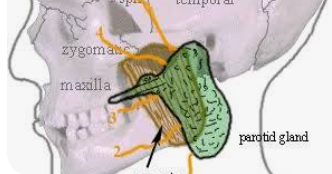
1 Where do the secretions of the parotid gland drain?The secretions of the parotid gland pass into the oral cavity via Stensons duct whose oral opening is opposite the second upper molar tooth.
2 Which structures pass through the parotid gland?
Facial nerve
External carotid artery
Retromandibular vein
Auriculotemporal nerve3 What is the lymphatic drainage of the parotid gland?
It contains lymph nodes within the substance of the gland itself. It then drains to the deep cervical nodes.
4 Which nerves supply the parotid gland?
Parasympathetic-Secretomotor
Sympathetic-Superior cervical ganglion
Sensory- Greater auricular nerve5 Outline where you would place the incision for a superficial parotidectomy.
The incision runs posterior to the mandible and up inferior to the tragus of the ear. Loss of cutaneous sensation to the ear lobe is therefore a risk of the procedure.

-
1/ Stenson's duct
2/ Main trunk of facial nerve, retromandibular vein, external carotid artery, auricular temporal nerve, parotid lymph nodes
3/ Superficial and deep parotid lymph nodes, which then empty into the superior deep cervical lymph nodes
4/ Facial nerve
5/ Modified Blair Incision -
1 Where do the secretions of the parotid gland drain?The secretions of the parotid gland pass into the oral cavity via Stensons duct whose oral opening is opposite the second upper molar tooth.
2 Which structures pass through the parotid gland?
Facial nerve
External carotid artery
Retromandibular vein
Auriculotemporal nerve3 What is the lymphatic drainage of the parotid gland?
It contains lymph nodes within the substance of the gland itself. It then drains to the deep cervical nodes.
4 Which nerves supply the parotid gland?
Parasympathetic-Secretomotor
Sympathetic-Superior cervical ganglion
Sensory- Greater auricular nerve5 Outline where you would place the incision for a superficial parotidectomy.
The incision runs posterior to the mandible and up inferior to the tragus of the ear. Loss of cutaneous sensation to the ear lobe is therefore a risk of the procedure.
@admin said in Station - parotid gland:
The incision runs posterior to the mandible and up inferior to the tragus of the ear. Loss of cutaneous sensation to the ear lobe is therefore a risk of the procedure
The incision is called Modified Blair and the loss of cutaneous sensation is due to the sacrifice/ injury to the great auricular nerve (a branch of the facial nerve)
-
1 Where do the secretions of the parotid gland drain?The secretions of the parotid gland pass into the oral cavity via Stensons duct whose oral opening is opposite the second upper molar tooth.
2 Which structures pass through the parotid gland?
Facial nerve
External carotid artery
Retromandibular vein
Auriculotemporal nerve3 What is the lymphatic drainage of the parotid gland?
It contains lymph nodes within the substance of the gland itself. It then drains to the deep cervical nodes.
4 Which nerves supply the parotid gland?
Parasympathetic-Secretomotor
Sympathetic-Superior cervical ganglion
Sensory- Greater auricular nerve5 Outline where you would place the incision for a superficial parotidectomy.
The incision runs posterior to the mandible and up inferior to the tragus of the ear. Loss of cutaneous sensation to the ear lobe is therefore a risk of the procedure.
@admin said in Station - parotid gland:
Parasympathetic
Parasympathetic (secretomotor) innervation
The parasympathetic supply increases the production of watery saliva. Its pathway is long and complex, beginning with the glossopharyngeal nerve (CN IX).- Origin: The preganglionic parasympathetic fibers arise from the inferior salivatory nucleus in the brainstem.
- Course: The fibers travel along the glossopharyngeal nerve and a small branch called the tympanic nerve, which passes through the middle ear.
- Synapse: The fibers continue as the lesser petrosal nerve and synapse in the otic ganglion, which is a collection of nerve cell bodies near the base of the skull.
- Supply: The postganglionic fibers then "hitchhike" along the auriculotemporal nerve (a branch of the trigeminal nerve) to reach and innervate the parotid gland. (Hence sometimes post parotidectomy when the great auricular nerve is sacrified or injured - it causes Frey's syndrome - where regenerating of parasympathetic fibers accidentally "rewire" themselves to connect with the sympathetic pathways that lead to the skin's sweat glands and blood vessels
Sympathetic innervation
The sympathetic supply reduces saliva production, causing a thicker, more viscous saliva via vasoconstriction.
• The postganglionic sympathetic fibers originate from the superior cervical ganglion and travel to the gland along the external carotid artery.Sensory innervation
The parotid gland receives its sensory supply from two nerves.
• Auriculotemporal nerve: This nerve provides general sensory innervation directly to the substance of the gland.
• Great auricular nerve: This nerve, a branch of the cervical plexus (C2 and C3), supplies the sensory innervation to the tough fascia or capsule of the parotid gland. -
A admin moved this topic from null on
-
A admin moved this topic from null on
-
A admin moved this topic from null on
Hello! It looks like you're interested in this conversation, but you don't have an account yet.
Getting fed up of having to scroll through the same posts each visit? When you register for an account, you'll always come back to exactly where you were before, and choose to be notified of new replies (either via email, or push notification). You'll also be able to save bookmarks and upvote posts to show your appreciation to other community members.
With your input, this post could be even better 💗
Register Login