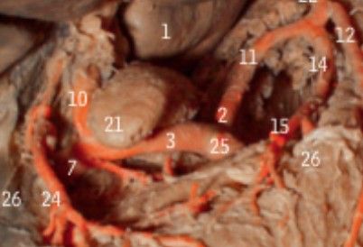
1: "Adductor longus",
2: "Arterial branch of vastus medialis",
3: "Corona of glans penis",
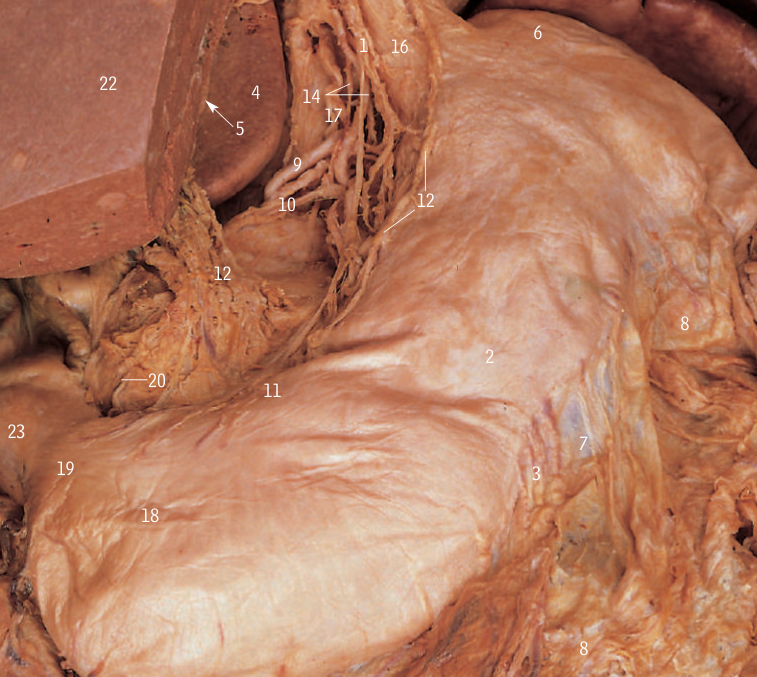
4: "External oblique aponeurosis",
5: "Fascia lata (cut edge)",
6: "Femoral artery",
7: "Femoral nerve",
8: "Femoral vein",
9: "Grachis",
10: "Great saphenous vein",
11: "Iliopsoas",
12: "Ilioitibal tract",
13: "Inguinal ligament",
14: "Nerve to vastus medialis",
15: "Pectineus",
16: "Perforating branch of profunda femoris artery",
17: "Rectus femoris",
18: "Saphenous nerve",
19: "Sartorius",
20: "Subartorial fascia (thickened aponeurosis)",
21: "Spermatic cord",
22: "Superficial circumflex iliac vein",
23: "Superficial epigastric vein",
24: "Superficial external pudendal vein",
25: "Superficial inguinal ring",
26: "Tensor fasciae latae deep to fascia lata",
27: "Valvular bulge lie in vein",
28: "Vastus lateralis",
29: "Vastus medialis",