Skip to content

Anatomy

39 Topics 94 Posts

This category can be followed from the open social web via the handle anatomy@isurg.org

  • isb7 Lower Limb

    Moved
    3
    1
    0 Votes
    3 Posts
    237 Views
    A
    Anterior [image: 1757607222780-090205a8-dc19-4036-baa6-c265e07a8591-image.png] Spoiler [image: 1757606977748-a7046645-fb1e-41d8-9a6a-60a411ec8218-image.png] Posterior [image: 1757606627233-8bb534ad-b401-4a07-8b0c-8ee290285be8-image.png] Spoiler[image: 1757606789866-c65988de-927e-49f3-a303-5fab5b16c349-image.png]
  • isb1 Skull foramina

    Moved
    5
    1
    0 Votes
    5 Posts
    450 Views
    A
    Station Topic: Brain Anatomy – Internal Carotid Artery Question (20 marks) [image: 1759597375048-2ab79662-441a-40de-b185-1c9d05240885-image.png] 
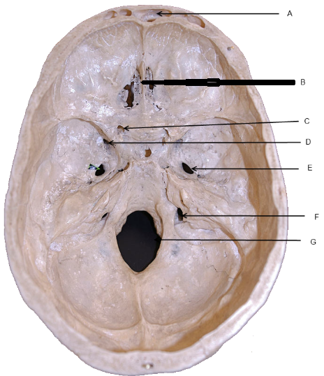
You are asked to demonstrate your knowledge of the internal carotid artery (ICA) and its relation to brain anatomy. Identify and describe the course of the internal carotid artery from the neck to the brain. (5 marks) Spoiler1 The ICA arises from the common carotid artery at the level of C3–C4 vertebrae. Cervical segment: Ascends vertically in the neck without branching. Petrous segment: Enters the carotid canal in the petrous temporal bone; runs anteromedially. Cavernous segment: Courses through the cavernous sinus; forms an S-shaped curve (the carotid siphon). Cerebral (supraclinoid) segment: Exits the cavernous sinus and pierces the dura mater at the roof of the cavernous sinus to enter the subarachnoid space; gives terminal branches to the brain. Tip: Remember mnemonic “Cervical, Petrous, Cavernous, Cerebral” to recall ICA segments. 2 List and explain the main branches of the internal carotid artery in the cranial cavity. (5 marks) SpoilerOphthalmic artery: First branch; supplies the orbit and optic nerve. Posterior communicating artery (PComm): Connects ICA to posterior cerebral artery; part of Circle of Willis. Anterior choroidal artery: Supplies choroid plexus, internal capsule, optic tract. Terminal branches: Anterior cerebral artery (ACA): Medial frontal and parietal lobes. Middle cerebral artery (MCA): Lateral convexity of cerebral hemisphere. Tip: ACA + MCA = terminal branches; remember PComm is part of collateral circulation. 3 Describe the areas of the brain supplied by these branches. (5 marks) SpoilerBranch Area Supplied Ophthalmic Eye, orbit, optic nerve Posterior communicating Connects ICA to posterior cerebral artery; collateral supply to occipital lobe Anterior choroidal Posterior limb of internal capsule, optic tract, globus pallidus, choroid plexus Anterior cerebral (ACA) Medial frontal and parietal lobes; leg motor/sensory cortex Middle cerebral (MCA) Lateral convexity of hemisphere; face and upper limb motor/sensory cortex, Broca/Wernicke areas Clinical Relevance of ICA (5 marks) Outline the clinical relevance of the internal carotid artery. Include at least two common pathologies and their implications. SpoilerAtherosclerosis / ICA stenosis: Can cause transient ischaemic attacks (TIAs) or stroke in MCA/ACA territories. Risk factors: hypertension, diabetes, smoking. Aneurysm formation: Common at bifurcation into MCA and ACA or posterior communicating artery. May cause subarachnoid haemorrhage or cranial nerve III palsy if PComm involved. Other considerations: SpoilerICA injury during carotid endarterectomy. Compression by tumours (e.g., pituitary adenoma in cavernous sinus) → ophthalmoplegia.
  • isb4 Humerus

    Moved
    3
    4
    0 Votes
    3 Posts
    129 Views
    A
    Right humerus lower end, A) Front B) Behind [image: 1760100719273-504353c4-52fc-4100-b8c5-d7b78397d7f0-image.png] 2 Capitulum. 5 Lateral epicondyle. 13 Trochlea. [image: 1760100719273-504353c4-52fc-4100-b8c5-d7b78397d7f0-image.png] 7 Medial epicondyle. 8 Medial supracondyle ridge 10 Olecranon fossa.
  • Larynx

    Moved
    1
    2
    0 Votes
    1 Posts
    159 Views
    No one has replied
  • Nerves to Mandible

    Moved
    1
    1
    0 Votes
    1 Posts
    50 Views
    No one has replied
  • Major haemorrhage protocol trauma station

    Moved
    4
    1
    0 Votes
    4 Posts
    178 Views
    A
    Explain the steps in Resuscitation and Monitoring (5 marks)
  • Station - parotid gland

    Moved
    4
    2
    0 Votes
    4 Posts
    157 Views
    S
    @admin said in Station - parotid gland: Parasympathetic Parasympathetic (secretomotor) innervation The parasympathetic supply increases the production of watery saliva. Its pathway is long and complex, beginning with the glossopharyngeal nerve (CN IX). Origin: The preganglionic parasympathetic fibers arise from the inferior salivatory nucleus in the brainstem. Course: The fibers travel along the glossopharyngeal nerve and a small branch called the tympanic nerve, which passes through the middle ear. Synapse: The fibers continue as the lesser petrosal nerve and synapse in the otic ganglion, which is a collection of nerve cell bodies near the base of the skull. Supply: The postganglionic fibers then "hitchhike" along the auriculotemporal nerve (a branch of the trigeminal nerve) to reach and innervate the parotid gland. (Hence sometimes post parotidectomy when the great auricular nerve is sacrified or injured - it causes Frey's syndrome - where regenerating of parasympathetic fibers accidentally "rewire" themselves to connect with the sympathetic pathways that lead to the skin's sweat glands and blood vessels Sympathetic innervation The sympathetic supply reduces saliva production, causing a thicker, more viscous saliva via vasoconstriction. • The postganglionic sympathetic fibers originate from the superior cervical ganglion and travel to the gland along the external carotid artery. Sensory innervation The parotid gland receives its sensory supply from two nerves. • Auriculotemporal nerve: This nerve provides general sensory innervation directly to the substance of the gland. • Great auricular nerve: This nerve, a branch of the cervical plexus (C2 and C3), supplies the sensory innervation to the tough fascia or capsule of the parotid gland.
  • Spermatic cord

    Moved
    1
    1
    0 Votes
    1 Posts
    96 Views
    No one has replied
  • The Larynx

    Moved
    1
    1
    0 Votes
    1 Posts
    61 Views
    No one has replied
  • isb6 Pelvis

    Moved
    3
    1
    0 Votes
    3 Posts
    291 Views
    A
    Identify 1-5 [image: 1745845181617-pelvi.png] 1Inguinal ligament 2Obturator membrane 3Anterior longitudinal ligament 4Iliolumbar ligament 5Lacunar ligament
  • Acute Pancreatitis

    Moved
    2
    0 Votes
    2 Posts
    181 Views
    A
    Differential Diagnosis Acute Pancreatitis Pancreatic collection Pancreatic pseudocyst Acute Cholecystitis Ascending cholangitis Cause of tachypnea ARDS Abdominal pain Pressure from Pseudocyst Sign of sepsis Respiratory complication such as pleural effusion Functions of Pancreas Endocrine -Alpha - glucagon -Beta - Insulin -Delta - Somatostatin Exocrine -Proteases -Lipolytic -Starch digestion Definition of acute pancreatitis Acute pancreatitis is a condition where the pancreas becomes inflamed (swollen) over a short period of time
  • Arcuate Line

    Moved
    1
    1
    0 Votes
    1 Posts
    250 Views
    No one has replied
  • The Pancreas

    Moved
    1
    0 Votes
    1 Posts
    143 Views
    No one has replied
  • The Liver

    Moved
    2
    1
    0 Votes
    2 Posts
    228 Views
    A
    Describe the contents of the porta hepatis. [image: 1739032208365-portahep.jpg] The porta hepatis is the gateway to the liver and contains several important structures: Common hepatic duct: Carries bile from the liver. Hepatic artery: Supplies oxygenated blood to the liver. Portal vein: Transports deoxygenated blood from the gastrointestinal tract and spleen to the liver. Autonomic nerve fibres: The sympathetic nerves originate from the coeliac axis, and parasympathetic fibres come from the vagus nerve. Lymphatic vessels and lymph nodes: Drain lymph from the liver. Explain the peritoneal attachments of the liver. The liver is connected to the abdominal wall and diaphragm by various ligaments: Right triangular ligament: A small fold of peritoneum between the upper and lower coronary ligaments at the posterior bare area of the liver. Falciform ligament: Runs from the umbilicus to the liver and carries the ligamentum teres. Left triangular ligament: Connects the posterosuperior part of the left lobe to the diaphragm and continues anteriorly as the left limb of the falciform ligament. Describe the hepatic veins and their drainage into the inferior vena cava (IVC). The hepatic veins are responsible for draining blood from the liver: Three main hepatic veins: Right, central, and left hepatic veins drain into the IVC. The central vein often drains into the IVC via the left hepatic vein, but in some individuals, it may drain directly into the IVC. Small hepatic venous tributaries: Drain blood directly from the liver's substance and enter the IVC more distally than the main veins. Zones of drainage: The zones correspond to the right, middle, and left thirds of the liver. The plane of the falciform ligament roughly demarcates the boundary between the left and middle zones. Outline the anatomical features of the biliary system. The biliary system includes the structures responsible for bile formation and transport: Hepatic ducts: The left and right hepatic ducts emerge from the liver and fuse at the porta hepatis to form the common hepatic duct. Cystic duct: Joins with the common hepatic duct to form the common bile duct (CBD). Common bile duct: Passes through the hepatoduodenal ligament and opens into the second segment of the duodenum. Pancreatic duct: The CBD joins the pancreatic duct at the ampulla of Vater, where the sphincter of Oddi controls the release of bile and pancreatic juices into the duodenum. Describe the anatomical features of the gallbladder. The gallbladder stores and concentrates bile: Location: Situated in the fossa between the right and quadrate lobes of the liver. Capacity: Approximately 50 mL of bile. Hartmann’s pouch: A small recess near the neck of the gallbladder where gallstones can form. Vascular supply: Receives blood from the cystic artery, which is a branch of the right hepatic artery. Venous drainage: Small veins drain into the right portal vein, as there is no distinct accompanying vein for the cystic artery. Histology: Primarily composed of mucosa lined by mucous-secreting columnar cells. The gallbladder wall also contains smooth muscle, which helps with bile contraction. Explain the embryological development of the gallbladder and its ducts. The development of the gallbladder and hepatic system begins early in fetal life: Liver and hepatic ducts: These structures form from a diverticulum of the ventral wall of the duodenum. This diverticulum differentiates into the liver and the hepatic ducts. Gallbladder and cystic duct: A second diverticulum forms from the side of the hepatic duct and differentiates into the gallbladder and cystic duct.
  • The Retroperitoneum

    Moved
    1
    0 Votes
    1 Posts
    98 Views
    No one has replied
  • The Greater and lesser sacs

    Moved
    1
    0 Votes
    1 Posts
    120 Views
    No one has replied
  • The small and large intestine

    Moved
    1
    0 Votes
    1 Posts
    90 Views
    No one has replied
  • The Stomach

    Moved
    1
    0 Votes
    1 Posts
    96 Views
    No one has replied
  • Anterior Abdominal Wall

    Moved
    1
    0 Votes
    1 Posts
    98 Views
    No one has replied