Mock4 - Foot and ankle
-
-
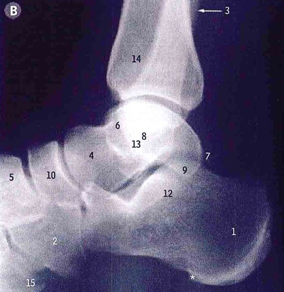
Name the tarsal bones
-
Articulation
The talus articulates:
with tibia and fibula
with navicular bone
with calcaneus
The navicular articulates:
with the three cuneiforms
with talus
The cuboid articulates:
with calcaneus
with lateral two metatarsals
The cuneiforms articulate:
with medial three metatarsals
with navicular bone
The calcaneus articulates:
with talus
with cuboid
Peroneus brevis
Avascular necrosis
-Posterior tibial artery supplies the body of the talus via:
Artery of tarsal canal (supplies most of talar body, dominant blood supply)
Deltoid branch supplies the medial portion of talar body
-Anterior tibial artery supplies head and neck of talus
-Peroneal artery supplies head and neck of talus via artery of tarsal sinusIn case of displaced talar neck fracture, which could lead to disruption of artery of tarsal canal, most of talar body will lose its blood supply (except medial portion which is supplied by the deltoid branch) and that will lead eventually to AVN
Anterior tibial artery
Loss of plantar flexion
Medial longitudinal arch
• Calcaneus, Talus, Navicular• 3 cuneiforms• 3 medial metatarsals
Lateral longitudinal arch
• Calcaneus• Cuboid• 2 lateral metatarsals
Transverse arch
• Cuboid, 3 cuneiforms• Bases of metatarsals
Mnemonic: Tom Has Very Nice Dogs & Pigs
Tibialis anterior
Extensor Hallucis longus
Anterior tibial vessels,
Anterior tibial nerve
Digitorum longus (Extensor)
Peroneus tertius -
-
Mnemonic: Tom Does Very Nice Hats
Tibialis posterior tendon
Flexor Digitorum longus
posterior tibial Vessels
posterior tibial Nerve
Hallucis longus (Flexor)Sustentaculum tali
Tibia
Fibula
TalusSynovial hinge joint
Plantarflexion
DorsiflexionAnkle joint is most stable in dorsiflexion, because the talus is wider anteriorly and narrower posteriorly.
(Note: In dorsiflexion, the wider anterior part of the talar trochlea wedges tightly into the mortise formed by the tibia and fibula.)
Talus and calcaneus
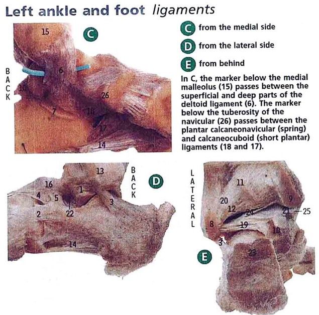
Inversion and eversion of footIdentify the Deltoid ligament, calcaneofibular ligament, posterior talofibular ligament:
Deltoid ligament (6)
Calcaneofibular ligament (3)
Posterior talofibular ligament (19)The dorsalis pedis pulse is found between the first two metatarsal bones.
The posterior tibial pulse is found 2cm-3cm below and behind the medial malleolus.The midtarsal joint, consisting of the talonavicular and the calcaneocuboid joints, is presumed to be responsible for the foot being both flexible and rigid during different parts of the stance phase of gait.
Hello! It looks like you're interested in this conversation, but you don't have an account yet.
Getting fed up of having to scroll through the same posts each visit? When you register for an account, you'll always come back to exactly where you were before, and choose to be notified of new replies (either via email, or push notification). You'll also be able to save bookmarks and upvote posts to show your appreciation to other community members.
With your input, this post could be even better 💗
Register Login