Mock 1 BCC
-
Stem 1: 78-year-old diabetic woman undergoing temporal artery biopsy, she has shoulder pain & a tender scalp.
When temporal artery biopsy was taken, a skin ulcer was found on her cheek.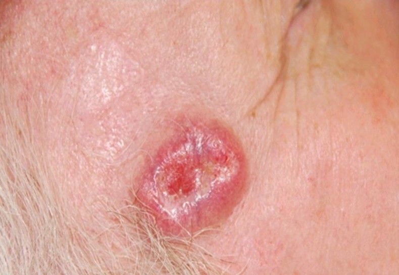
Describe what you see?Pearly papule
Central ulcer
Granulation tissue on base
Inverted edges
Surrounding telangiectasiaWhat is your differential diagnosis?
BCC
SCC
TB
Actinic keratosisNatural Hx of BCC?
Indolent with slow progression
Locally destructive
but limited potential to metastasizeExaminer gives you histology report. What will you look for in the report?
Deep margin involvement (i.e. clear margin or not)
Size and InvasionIf unclear margins, what to do?
Re-excision with safety margin, with frozen section or MOHs micrographics surgery.
If there is lymph node involvement, what to do?
Lymph nodes biopsy (FNAC or excisional biopsy)
What is frozen section?
It is a laboratory procedure that allows for a quick microscopic examination of tissue to help diagnose disease
What are advantages of frozen section?
Give rapid intraoperative pathological diagnosis
Can you explain the steps?
The surgeon takes a small piece from a tissue or tumour and send it for analysis
The pathologist freezes it and section it and immediately cut it
The section will be stained and reported immediately when the results come outHow do malignant cells reach lymph nodes?
Cancer cells and the secrete growth factors which are taken up by the lymphatic capillaries and transported towards the LN.
These growth factors induce lymph-angiogenesis thereby promoting LN metastasisSkin graft was done, and the patient had graft failure subsequently. What are the possible causes for graft failure?
Wound infection
Hematoma
RejectionCommon organisms for wound infection?
Staph. aureus.
Streptococcus.What to do to diagnose it?
Swab for C&S
Blood test (CRP, WCC)What is MRSA?
Methicillin-resistant Staphylococcus aureus.
What to do if swab for MRSA is positive?
Isolation of patient
Inform infection control team and follow infection control protocol
Wound debridement. I&D if abscess
Give antibiotics according to trust/local protocol / microbiology adviceHow to decolonise MRSA in a carrier patient (not infected)?
With mupirocin nasal
Chlorhexidine body wash / shampooWho will you involve in the management?
Ward nurse
Microbiologist
infection control teamPatient developed axillary LN enlargement. LN biopsy showed large cells with bilobed nucleus with prominent eosinophilic inclusion with nuclei resembling an owl's eye appearance.
What is your diagnosis now?Reed-Sternberg cells (owl eye appearance) therefor Hodgkin's lymphoma
Define carcinoma?
Is a type of cancer that develops from epithelial cells
Features of malignant cells?
Increased number of mitotic figures
Abnormal mitoses
Hyperchromatism (nuclei are darkened due to increased concentration of DNA)
Pleomorphism (varied size and shape of cell)
Increased nuclear to cytoplasmic ratio -
Stem2: 78-year-old diabetic woman referred for temporal artery biopsy, having shoulder pain and tender scan for one week. When performing a temporal artery biopsy, you find nodular lesion with some ulcerations in the cheek of the patient suggestive of primary skin carcinoma probably Basal cell carcinoma.
Why is temporal artery being biopsy in this case?
To test of presence of giant cell arthritis.
What is the usual medical treatment of this condition which is started before the biopsy?
Large dose of corticosteroid.
Why is steroid therapy often started before biopsy is taken?
To protect vision from ophthalmic artery insolvent from arthritis.
What 4 items of data would you want to see in skin pathology summary report?
The type of tumour verification.
Tumor diameter.
Tumor thickness.
Margin clearance, lateral and deep margin.What features of that tumour may give insight into the risk of metastatic disease?
Presence or absent blood or lymphatic invasion.
Six months later the patient readmitted following a mechanical fall in the community. Radiology revealed a fracture neck of femur, requiring surgical intervention.
What factors in the scenario can predisposed to this osteoporotic fracture?Female.
Age and post menopause.
Steroid.From the biochemical perspective, what do you expect the serum calcium and phosphate levels to be, considering this patient is osteoporotic?
Both of them will be normal.
During surgery, the bone matrix appears to be soft and fragile. A sample sent to the histology and revealed plasmacytoma or myeloma. What will you find in this?
Neuroplastic, monoclonal, perforations of plasma cell.
What may such tumours commonly secrete into the serum?
Myeloma, immunoglobulins or fragmentation of immunoglobulins.
What is Bence Jones protein?
A light chain or paraprotein, which may be identified in urine.
Apart from osteoporosis, what may be the cause of fracture of this patient?
Tumour itself.
Bone lysis by the tumour.The screw is required to repair the fracture and some radiotherapy is planned. However, the wound decide heals poorly with a septic breakdown. What 4 factors likely to contribute wound sepsis?
Steroid.
Diabetic.
Immunocompromise because plasmacytoma.
Foreign body processes.Despite antibiotic treatment, the hip infection persists; excision of the hip, and the fracture site repair followed by a full hip replacement. Patient arrested 24 hours after surgery.
What is the cause of this?Venous thromboembolism
Hello! It looks like you're interested in this conversation, but you don't have an account yet.
Getting fed up of having to scroll through the same posts each visit? When you register for an account, you'll always come back to exactly where you were before, and choose to be notified of new replies (either via email, or push notification). You'll also be able to save bookmarks and upvote posts to show your appreciation to other community members.
With your input, this post could be even better 💗
Register Login