-
What is the remnant of the left umbilical vein in the adult liver, and where is it located?
Answer: Ligamentum teres hepatis, found in the anterior left fossa.
it is also known as the round ligament of the liver, is a fibrous band that connects the liver to the umbilicus. It's a remnant of the umbilical vein, which carried blood from the placenta to the liver during fetal development.Which hepatic structure is the remnant of the fetal ductus venosus, and what is its function in the fetal circulation?
Answer: Ligamentum venosum, the fibrous remnant of the ductus venosus, which functioned as a shunt for oxygenated blood from the left umbilical vein to the inferior vena cava.Through which anatomical plane does the functional division of the liver occur?
Answer: Through the fossae of the gallbladder and inferior vena cava (IVC).Name the three main hepatic veins and describe their drainage pathways.
Answer:
Right hepatic vein
Left hepatic vein
Central hepatic vein
The central hepatic vein usually drains into the left hepatic vein near its termination but may drain directly to the inferior vena cava (IVC). The right and left hepatic veins drain directly into the IVC.What are the boundaries of Calot’s triangle and which structures are contained within it?
Answer: The liver, the cystic duct, and the common hepatic duct.Where is Hartmann’s pouch typically located, and what is its clinical significance?
Answer: Hartmann’s pouch is located on the ventral aspect of the gallbladder, just proximal to the neck. It is a potential site for gallstone blockage. -
Describe the contents of the porta hepatis.
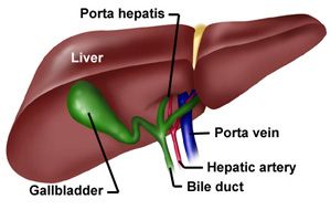
The porta hepatis is the gateway to the liver and contains several important structures:Common hepatic duct: Carries bile from the liver.
Hepatic artery: Supplies oxygenated blood to the liver.
Portal vein: Transports deoxygenated blood from the gastrointestinal tract and spleen to the liver.
Autonomic nerve fibres: The sympathetic nerves originate from the coeliac axis, and parasympathetic fibres come from the vagus nerve.
Lymphatic vessels and lymph nodes: Drain lymph from the liver.Explain the peritoneal attachments of the liver.
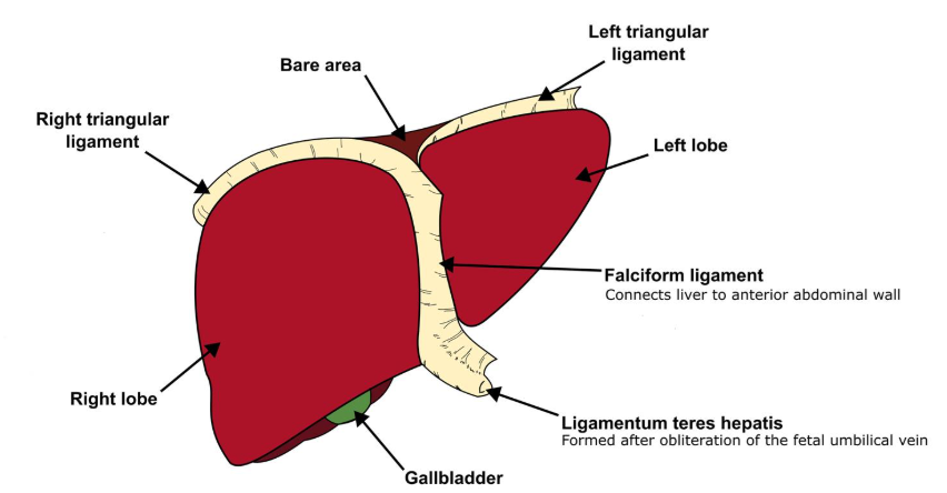
The liver is connected to the abdominal wall and diaphragm by various ligaments:
Right triangular ligament: A small fold of peritoneum between the upper and lower coronary ligaments at the posterior bare area of the liver.
Falciform ligament: Runs from the umbilicus to the liver and carries the ligamentum teres.
Left triangular ligament: Connects the posterosuperior part of the left lobe to the diaphragm and continues anteriorly as the left limb of the falciform ligament.Describe the hepatic veins and their drainage into the inferior vena cava (IVC).
The hepatic veins are responsible for draining blood from the liver:
Three main hepatic veins: Right, central, and left hepatic veins drain into the IVC. The central vein often drains into the IVC via the left hepatic vein, but in some individuals, it may drain directly into the IVC.
Small hepatic venous tributaries: Drain blood directly from the liver's substance and enter the IVC more distally than the main veins.
Zones of drainage: The zones correspond to the right, middle, and left thirds of the liver. The plane of the falciform ligament roughly demarcates the boundary between the left and middle zones.Outline the anatomical features of the biliary system.
The biliary system includes the structures responsible for bile formation and transport:
Hepatic ducts: The left and right hepatic ducts emerge from the liver and fuse at the porta hepatis to form the common hepatic duct.
Cystic duct: Joins with the common hepatic duct to form the common bile duct (CBD).
Common bile duct: Passes through the hepatoduodenal ligament and opens into the second segment of the duodenum.
Pancreatic duct: The CBD joins the pancreatic duct at the ampulla of Vater, where the sphincter of Oddi controls the release of bile and pancreatic juices into the duodenum.Describe the anatomical features of the gallbladder.
The gallbladder stores and concentrates bile:
Location: Situated in the fossa between the right and quadrate lobes of the liver.
Capacity: Approximately 50 mL of bile.
Hartmann’s pouch: A small recess near the neck of the gallbladder where gallstones can form.
Vascular supply: Receives blood from the cystic artery, which is a branch of the right hepatic artery.
Venous drainage: Small veins drain into the right portal vein, as there is no distinct accompanying vein for the cystic artery.
Histology: Primarily composed of mucosa lined by mucous-secreting columnar cells. The gallbladder wall also contains smooth muscle, which helps with bile contraction.Explain the embryological development of the gallbladder and its ducts.
The development of the gallbladder and hepatic system begins early in fetal life:
Liver and hepatic ducts: These structures form from a diverticulum of the ventral wall of the duodenum. This diverticulum differentiates into the liver and the hepatic ducts.
Gallbladder and cystic duct: A second diverticulum forms from the side of the hepatic duct and differentiates into the gallbladder and cystic duct. -
A admin moved this topic from Anatomy on
-
A admin moved this topic from on
Hello! It looks like you're interested in this conversation, but you don't have an account yet.
Getting fed up of having to scroll through the same posts each visit? When you register for an account, you'll always come back to exactly where you were before, and choose to be notified of new replies (either via email, or push notification). You'll also be able to save bookmarks and upvote posts to show your appreciation to other community members.
With your input, this post could be even better 💗
Register Login