Sinuses
-
Examiner Mark Scheme (20 marks total)
- Definition & General Features (3 marks)
Endothelium-lined venous channels located between periosteal and meningeal layers of dura mater (1)
Valveless system allowing bidirectional blood flow (1)
Drain venous blood from brain, skull, meninges, and CSF via arachnoid granulations (1)- Major Sinuses Identified (6 marks)
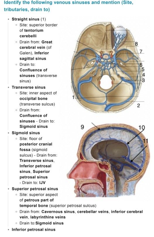
Superior sagittal sinus
Inferior sagittal sinus
Straight sinus
Transverse sinuses
Sigmoid sinuses
Cavernous sinuses
(Allow alternatives such as occipital sinus, intercavernous sinuses)- Anatomical Course & Drainage (6 marks)
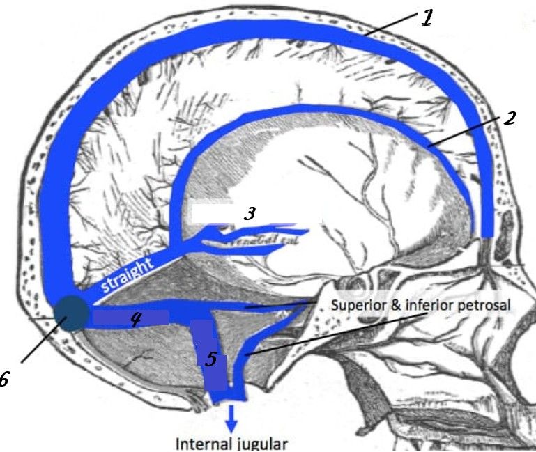
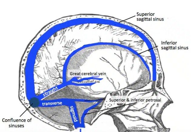
Superior sagittal sinus: runs in falx cerebri → drains to confluence of sinuses (1)
Inferior sagittal sinus: joins great cerebral vein → forms straight sinus (1)
Straight sinus: drains to confluence (1)
Transverse sinuses: from confluence → become sigmoid sinuses (1)
Sigmoid sinuses: drain into internal jugular vein (1)
Cavernous sinus: drains via superior & inferior petrosal sinuses (1)- Cavernous Sinus Detail (3 marks)
Location: either side of sella turcica (1)
Contents:
Internal carotid artery
CN III, IV, V1, V2, VI (1)
Connections: facial vein via superior ophthalmic vein (1)- Clinical Relevance (2 marks)
Cavernous sinus thrombosis (spread from facial infections) (1)
Raised intracranial pressure affecting venous drainage / CSF absorption (1)
Examiner Prompts (if candidate struggling)
“What structures are related to the cavernous sinus?”
“Where does venous blood ultimately drain?”
“Why are infections of the face dangerous?”
Confluence of sinuses = meeting point (superior sagittal, straight, occipital)
Internal jugular vein is final drainage pathway
“Danger area of face” → cavernous sinus via valveless veins
CN VI most vulnerable in cavernous sinus pathologyModel Candidate Answer (Concise)
“The dural venous sinuses are valveless, endothelium-lined channels between layers of dura that drain venous blood and CSF from the brain. The major sinuses include the superior and inferior sagittal, straight, transverse, sigmoid, and cavernous sinuses. Blood flows from cortical veins into the superior sagittal sinus, through the confluence, into transverse and sigmoid sinuses, and finally into the internal jugular vein.
The cavernous sinus lies beside the sella turcica and contains the internal carotid artery and cranial nerves III, IV, V1, V2, and VI. It communicates with the facial vein, making it clinically important in cavernous sinus thrombosis. Dysfunction of these sinuses can also impair CSF absorption and raise intracranial pressure.”
Hello! It looks like you're interested in this conversation, but you don't have an account yet.
Getting fed up of having to scroll through the same posts each visit? When you register for an account, you'll always come back to exactly where you were before, and choose to be notified of new replies (either via email, or push notification). You'll also be able to save bookmarks and upvote posts to show your appreciation to other community members.
With your input, this post could be even better 💗
Register Login