Anatomy station Transpyloric plane
-
MRCS OSCE: Abdomen Station (L1 Cross-Section)
Question 1: Demonstrate the transpyloric plane on this subject.
Model Answer:
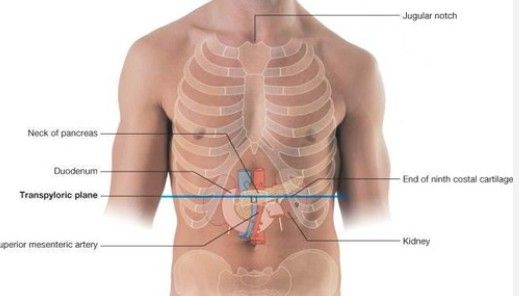
The transpyloric plane (Addison’s plane) is a horizontal plane passing through the L1 vertebral body. It is found:Surface Marking: Midway between the jugular notch and the upper border of the symphysis pubis.
Alternatively: Approximately a hand’s breadth below the xiphisternal joint, or midway between the xiphoid process and the umbilicus.
Question 2: Is this axial cut section viewed from below (inferiorly) or above (superiorly)?
Model Answer:
Radiological and anatomical cross-sections are traditionally viewed from below (looking upward).Clinical Correlation: This matches the orientation of a CT scan, where the patient’s right is on the left side of the image.
Question 3: Identify the space between the anterior abdominal wall and the liver, and the space behind the stomach.
Model Answer:Space between wall and liver: The Subphrenic space (specifically the right or left anterior subphrenic space).
Space behind the stomach: The Lesser Sac (Omental Bursa).
Question 4: Describe the blood supply of the stomach.
Model Answer:
The stomach is supplied by branches of the Celiac Trunk:Lesser Curvature: Left gastric artery (from celiac) and Right gastric artery (from common hepatic).
Greater Curvature: Left gastro-omental (gastropiploic) artery (from splenic) and Right gastro-omental artery (from gastroduodenal).
Fundus: Short gastric arteries (from splenic).
Question 5: Locate the gallbladder on this subject.
Model Answer:
The gallbladder is located at the point where the lateral border of the rectus abdominis (linea semilunaris) intersects the 9th costal cartilage.Question 6: If you move your finger down along the costal margin to the mid-axillary line, what organ is underneath?
Model Answer:
The Spleen.Anatomical context: The spleen lies deep to the 9th, 10th, and 11th ribs on the left side, extending as far forward as the mid-axillary line. (Note: If the examiner is referring to the right side, the answer would be the Liver/Right Kidney, but the "costal cartilage" prompt in MRCS usually refers to the splenic clinical examination).
Summary of L1 Structures (The "Everything in the Picture" Question)
If the examiner asks you to name everything visible at the L1 level, ensure you mention:Pylorus of the stomach.
Hilus of both kidneys (the left is slightly higher than the right).
Fundus of the gallbladder.
Neck of the pancreas.
Origin of the Superior Mesenteric Artery (SMA) from the Aorta.
Conus medullaris (termination of the spinal cord).
Hello! It looks like you're interested in this conversation, but you don't have an account yet.
Getting fed up of having to scroll through the same posts each visit? When you register for an account, you'll always come back to exactly where you were before, and choose to be notified of new replies (either via email, or push notification). You'll also be able to save bookmarks and upvote posts to show your appreciation to other community members.
With your input, this post could be even better 💗
Register Login