Vascular
-
Deep Vein Thrombosis
You are currently a senior surgical doctor on call. Your next patient, Tom (61-year-old male) complaining of leg pain. Please take a history and carry out a relevant examination.
Patient History:
Tom, 61 y/o, recently retired – used to work as a business analyst.
You have been experiencing increasing leg pain for the past few days. The pain is located only at the left calf (only answer when specifically asked about the location). Pain is gradually getting worse and does not radiate to other parts of the body. Touching it makes it worse, and resting makes it better. The skin around the left calf feels hot and red; otherwise, no other skin changes. No previous injury towards calves.
There’s no coughing/ haemoptysis/ breathlessness. No fever/weight loss/ night sweats.
Ideas, Concerns, Expectations:
You think it might just be heart failure as you have been experiencing lower leg swelling for the past few years – which is why you are on furosemide. You can be concerned about the pain as there's limiting your rehab from your hip replacement which you had 1-2 weeks ago – you want to get better as soon as possible. You want to find out what is going on and have some painkillers.
Past Medical History:
Obesity, Diabetes Type 2, Hypertension, Heart Failure.
No previous malignancy with treatment within 6 months/ palliative.
No previous history of DVT/ PE.
Past Surgical History:
You just received a hip replacement 1-2 weeks ago, and due to pain, you have been quite immobile lately. Have been resting on a chair most of the time.
Drug History:
Metformin, Amlodipine, Multi-vitamins, Atorvastatin, Furosemide
Allergic to penicillin (anaphylactic reaction)
Family History:
Nil
Social History:
Do not smoke (used to smoke 10 cigarettes a day for 15 years)
Drink a beer every night with dinner
Live with wife
Retired - used to work as a business analyst.
Examination Findings:
Left calf erythema and warmth to touch
Distension of superficial veins and tenderness on palpitation of the left calf
Mild pitting oedema and swelling on both calves
The left calf swelling is worse than the right
Chest clear on auscultation
Differentials:
Deep Vein Thrombosis
Cellulitis
Superficial thrombophlebitis
Heart Failure
Trauma including calf muscle haematoma, calf muscle tear/ Achilles' tendon tear
Investigations:
Bedside:
Observations
ECG (sinus tachycardia / S1 Q3 T3 changes in PE)
Calculate Well's Score for DVT
Bloods:
Anticoagulation screen
Routine blood tests: FBC, Bone profile LFTs, U&Es, CRP (infections/renal function for scans and treatment)
Consider D-Dimer
Consider Antiphospholipid antibodies (if unprovoked)
Imaging:
USS scan (if Well’s score at least 2 points and is within 4 hours from onset)
CXR/ CT pulmonary angiogram (if suspecting pulmonary embolism)
Management:
Conservative:
Control risk factors: obesity, heart failure, smoking.
Medical:
Confirmed/suspected proximal DVT: apixaban/ rivaroxaban (first-line).
Alternative: LMWH (at least 5 days followed by dabigatran/edoxaban/DOAC)
Anticoagulation treatment for at least 3 months with confirmed proximal DVT/PE.
Consider LMWH/unfractionated heparin instead of apixaban/rivaroxaban – if low renal function
LMWH and a vitamin K antagonist (VKA) for at least 5 days in phospholipid syndrome
Follow-up from the anticoagulation team
Viva Questions:
What are the risk factors for VTE?
Immobility: Prolonged bed rest, long flights, or extended periods of sitting can increase the risk.
Surgery or Trauma: Any surgery, especially orthopedic or major surgeries, increases the risk. Trauma or injury can also raise the likelihood of VTE.
Cancer: Certain cancers and cancer treatments elevate the risk of blood clot formation.
Age: Risk increases with age, especially over 60.
Obesity: Higher body mass index (BMI) is associated with increased risk.
Pregnancy and Postpartum: Pregnancy and the postpartum period increase the risk due to changes in blood flow and hormonal fluctuations.
Family History: Having a family history of blood clots or inherited clotting disorders can raise the risk.
Hormonal Medications: Use of birth control pills or hormone replacement therapy can increase the risk.
Medical Conditions: Conditions like heart disease, lung disease, and inflammatory disorders may increase the risk of VTE.
Smoking: Smoking can contribute to blood clot formation.
Previous History of VTE: Individuals with a history of previous blood clots are at a higher risk of recurrence.
How do you calculate the Well’s score for DVT?
The criteria and corresponding points in the Wells' score for DVT are as follows:
Active cancer (treatment within the last 6 months or palliative): 1 point
Calf swelling >3cm compared with the asymptomatic side: 1 point
Collateral non-varicose superficial veins (in the symptomatic leg): 1 point
Entire leg swelling: 1 point
Pitting edema confined to the symptomatic leg: 1 point
Paralysis, paresis, or recent cast immobilization of the lower extremities: 1 point
Recently bedridden for more than three days or major surgery within the last four weeks: 1 point
Localised tenderness along the deep venous system: 1.5 points
Previously documented DVT: 1.5 points
The total points are used to categorize the probability of a patient having DVT:
Score of 0 or less: Low probability of DVT
Score of 1-2: Moderate probability
Score of 2 or higher: High probability
What are the complications of DVT?
Pulmonary Embolism (PE): A life-threatening condition where a blood clot travels to the lungs.
Post-thrombotic Syndrome (PTS): Long-term complications, including chronic pain and swelling in the affected leg.
Chronic Venous Insufficiency (CVI): Damage to vein valves leading to reduced blood flow, causing symptoms like swelling and skin changes.
Recurrent DVT: Increased risk of experiencing another episode of DVT.
Venous Ulcers: Severe cases of CVI can lead to open sores or ulcers on the skin.
-
Peripheral Vascular Disease
Doctor Instruction:
You are currently a senior surgical doctor on call. Your next patient is Larry, a 61-year-old man with leg pain. Please take a history and perform a relevant examination.
Patient History:
Larry, 61 y/o M, retired engineer.
About 7 months ago, you started having pain in both legs that seemed triggered by walking. The pain feels like a cramp and most intensely in your calves and thighs.
You love going on long walks with your wife on a route that winds through a hilly forest. Because of the pain, however, you are forced to take a rest break every 100 metres, and this is getting worse. The pain is not worse or better when walking up or down a hill.
If you are specifically asked, share that you have been having trouble ‘getting it up’ in the bedroom.
You have not noticed any back pain, joint pains, or stiffness in your legs or locking or giving way of your knee joints.
Ideas, Concerns, Expectations:
You think that the pain could be a normal part of the ageing process, but it bothers you that it has been having such a big impact on supposedly the most enjoyable part of your day - spending time with your wife on nice walks. The pain also makes you feel like a burden when your son or grandkids join you on your walk. You would like a treatment plan that works as quickly as possible and do not mind if it involves taking medication.
Past Medical History:
Hypertension, 2 previous myocardial infarctions – 2 and 5 years previously, benign prostatic hyperplasia
Drug History:
Low-dose daily clopidogrel, Propranolol, Atorvastatin, amlodipine, ramipril, indapamide, tamsulosin (No known drug allergies)
Family History:
Sister had a leg clot when she was pregnant.
Social History:
Smoker – 40 cigarettes a day for 30 years.
You drink an occasional pint of beer on the weekend.
No recreational drug use.
You live with your wife at home.
You used to do most of the cooking and cleaning but have lately handed over your household responsibilities to your wife because of your poor health.
You can just about manage the stairs in your home, but you wish you could move into a single-storey flat as it would make your life more convenient.
Examination Findings:
An examination of the peripheral vasculature is most appropriate.
The patient exhibits a large body habitus.
There is an absence of hair on the lower legs.
Both legs look pale and feel cold on palpation.
Capillary refill times are less than 2 seconds bilaterally.
The right dorsalis pedis pulse is present on palpation.
The left dorsalis pedis and both posterior tibial pulses are absent on palpation.
Buerger’s test is positive bilaterally.
Differentials:
Peripheral vascular disease
Spinal canal stenosis
Musculoskeletal causes
Nerve root pain
To rule out DVT
The history and examination above are a classic presentation of intermittent claudication of peripheral arterial disease. Patients with peripheral arterial disease can experience calf pain that is exacerbated by walking and relieved by rest. Typical progression will see the distance a patient can walk without resting (aka the 'claudication distance') decrease over time until the rest pain of critical limb ischaemia is reached.
Additionally, Mr Done is exhibiting signs of aortoiliac involvement: erectile dysfunction and pain in his thighs.
Another key differential of bilateral calf pain exacerbated by walking is the 'neurogenic claudication' caused by spinal canal stenosis. This typically presents as a bilateral posterior leg pain that is relieved by spine flexion.
Investigations:
Bedside:
Blood pressure (hypertension is a modifiable cardiovascular risk factor)
Scoring systems: Rutherford Classification, Fontaine Classification
Doppler US (can confirm the presence or absence of foot pulses when the pulse cannot be felt on palpation)
Ankle brachial pressure index:
A value of 0.5-0.9 is indicative of peripheral arterial disease.
Values of <0.5 indicate critical limb ischaemia.
A normal ABPI does not rule out peripheral arterial disease.
Diabetic or calcified vessels may record ABPI values higher than 0.9 even if peripheral arterial disease is present.
Bloods:
HbA1c and blood glucose
Lipid profile
Coagulation Screen
FBC
Imaging:
Duplex ultrasound is conducted before any revascularisation procedure
CT/MR angiography
Management:
Conservative:
Lifestyle intervention to target risk factors:
Stop smoking
Cut down on alcohol, targeting <14 units per week
Eat a balanced diet
Decrease BMI
Supervised or unsupervised exercise programme:
Exercise programmes and lifestyle interventions are the first-line treatment for peripheral arterial disease.
Further treatment with medical interventions like Naftidofuryl oxalate or surgical and procedural interventions should only be considered if there is insufficient symptomatic improvement with the conservative treatment above.
Medical:
Secondary prevention of cardiovascular disease:
Antiplatelet (Clopidogrel 75mg daily)
Statin (Atorvastatin 80mg daily)
Good control of diabetes and hypertension.
Naftidrofuryl oxalate (naftidrofuryl oxalate can be prescribed to provide symptomatic relief for patients who are unfit for or decline surgical or procedural intervention)
Surgical or procedural:
Interventional – angioplasty and stenting
Surgical – bypass surgery (a last-resort intervention considered after angioplasty)
Viva Questions:
What are the risk factors for the development of peripheral arterial disease?
Smoking: Significant contributor to artery damage.
Diabetes: High blood sugar damages blood vessels.
High Blood Pressure: Contributes to artery narrowing.
High Cholesterol: Increases plaque buildup in arteries.
Age: Risk increases after 50.
Obesity: Adds strain to the circulatory system.
Family History: Genetic predisposition.
Inactivity: Lack of physical movement impacts circulation.
Cardiovascular Issues: Prior heart disease or stroke.
High Homocysteine: Elevates atherosclerosis risk.
Chronic Kidney Disease: Increases PAD risk.
Inflammatory Conditions: Such as rheumatoid arthritis can raise the risk.
What are the complications of peripheral arterial disease?
Critical Limb Ischemia (CLI): Severe reduction in blood flow to limbs, leading to ulcers, sores, and potential amputation.
Pain and Discomfort: Leg pain during activity due to reduced blood flow.
Wound Healing Issues: Slow healing of leg and foot ulcers.
Infections: Increased risk due to poor blood circulation.
Amputation: In severe cases, limb amputation may be necessary.
Cardiovascular Complications: Higher risk of heart attack and stroke.
Reduced Quality of Life: Limited mobility impacts daily activities and quality of life.
What are the clinical features seen in developing critical limb ischaemia or acute limb ischaemia?
Sudden Onset: Symptoms appear suddenly and progress rapidly, requiring urgent medical attention.
Severe Pain: Intense, persistent pain, especially at rest, not relieved by typical pain-relief methods.
Pulse Absence: Reduced or absent arterial pulses in the affected limb.
Skin Changes: Pale, discolored skin and a cooler limb due to poor circulation.
Numbness or Tingling: Sensations of numbness or tingling.
Muscle Weakness: Potential weakness or paralysis in the affected limb.
Ulcers or Gangrene: Non-healing sores or signs of tissue death (gangrene) due to inadequate blood supply.
-
Abdominal Aortic Aneurysm (AAA)
Doctor Instruction:
You are currently a senior surgical doctor on call. Your next patient is 64-year-old Harry complaining of abdominal discomfort. Please take a history and perform a relevant examination.
Patient History:
You are Harry - a 64-year-old male - retired construction worker.
You've had some vague aching pain in the middle part of your upper abdomen which comes and goes. Not worsened by eating/moving. It is gradually getting worse. It radiates to the back. You'd rate the pain 4/10 but has got worse. Sometimes you feel lightheaded upon standing.
Bowels are normal, no urinary symptoms, no vomiting, no heartburn. Never had screening tests. No fever, no weight loss, no blood in stool, no loss of consciousness.
You haven't had any fevers, weight loss or black stool. You sometimes feel a bit faint, especially when standing up. No collapse. No loss of consciousness.
Ideas, Concerns, Expectations:
You have no idea what this is - possibly maybe reflux. You're not really worried about it, but you hope to just find out what is going on - as told by your wife.
Past Medical History:
Hypertension
High cholesterol
Angina
Type 2 diabetes.
Drug History:
Amlodipine, ramipril, GTN spray, bisoprolol, insulin, atorvastatin
NKDA
Family History:
Your father died of bowel cancer at the age of 70.
Your mother has diabetes.
Social History:
You live at home with your wife and frequently take care of your mother.
You've smoked 20 cigarettes a day since you were 15.
You used to binge drink when you were younger, but nowadays, you'll drink the occasional six or so cans of lager at night.
You're a retired construction worker.
Examination Findings:
Observations are normal
Overweight body habitus
Nicotine staining on right hand
Corneal arcus
Epigastric tenderness
No organomegaly
Strong Pulsatile and expansile central abdominal mass
Differentials:
Abdominal aortic aneurysm - Central abdominal pain, pulsatile mass, male, age, radiates to back, vascular history, smoker.
Pancreatitis (alcohol history) - Epigastric pain, but may have indication of exocrine dysfunction (E.g. steatorrhoea), and it wouldn't be pulsitile
GORD - Would have retrosternal burning pain, worse with classic triggers.
Constipation/obstruction - May be faecal mass (but wouldn't be pulsitile) Passed stool today, no change in bowel habit
Diverticulitis - Usually LLQ pain, fever, no pulsatile masses
Investigations:
Bedside:
Observations (hypotension and tachycardia in rupture)
ECG (cardiovascular co-morbidities)
Bloods:
FBC, U&E, CRP, LFT
Amylase - Assess for acute pancreatitis
Coagulation screen - Risk of bleeding
Imaging:
Abdo USS (diagnostic for AAA and look for gallstones / dilated biliary tree)
CT angiogram (guides elective surgery for repair for AAA and to diagnose/exclude ruptured AAA in haemodynamically stable patients)
CT Abdomen (pancreatitis)
Specialist Test:
Consider OGD (if suspecting upper GI cause)
Data Interpretation:
Please interpret Image 1, once interpreted, move on to Image 2 below:
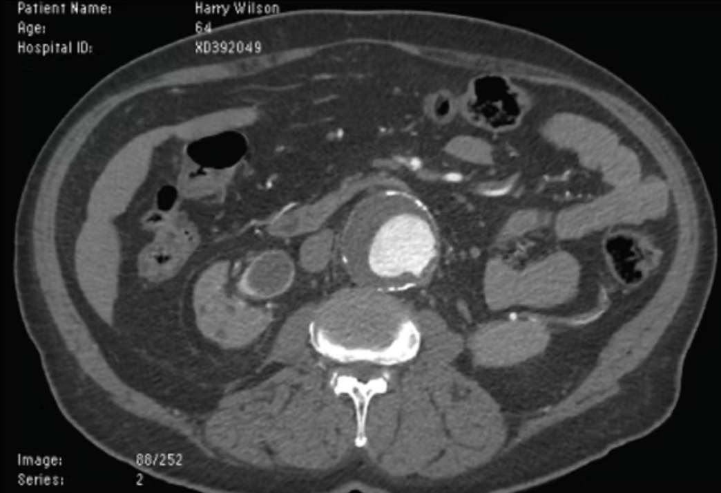
Candidate should confirm that its the right patient Name & Age and mention they would check if the patient ID is correct.
CT scan shows adequate contrast enhancement of the aorta with a clear view of an aneurysm. The aorta is calcified around. There is no leakage of blood from the aneurysm. There is no sign of a hyperdense crescent (which would have indicated a thrombus and impending rupture).

Interpretation
Image shows that the measurement of the AAA 5.5 cm, indicating the need for a vascular referral for an elective endovascular repair.
This does not change the appropriate management but further confirms it as being symptomatic is also a reason for referral for intervention.Management:
Conservative:
Patient Education
Contact DVLA if >6cm AAA
Stop driving if AAA is above 6.5cm
Stricter rules apply to drivers of heavy vehicles e.g. lorry drivers/ bus
Life style changes: smoking cessation, healthy diet and exercise
Medical:
Manage risk factors e.g. hypertension, hyperlipidaemia, diabetes
Surgical:
Refer to the vascular team if the diameter of the aorta is above 3cm. Urgent referral if more than 5.5cm
Offer elective repair in patients with symptomatic aneurysm, aneurysm diameter above 5.5cm or diameter growing >1cm per year
Methods: endovascular aneurysm repair (EVAR) / Open repair via a laparotomy
Screening:
All men in England are offered an USS scan at age 65 to detect asymptomatic AAA. Can be considered in women at age of 70 with risk factors such as smoking, hyperlipidemia, high blood pressure, COPD, cardiovascular disease, and family history.
Yearly screening in patients with aneurysms 3-4.4cm
Three monthly screening in patients with aneurysms 4.5-5.4cm
Ruptured AAA:
Emergency Surgery (patient to be urgently transferred to the theatre)
Permissive hypotension (aiming lower than normal blood pressure)
Surgical repair should not be delayed by diagnostic imaging
For patients with co-morbidities and poor prognosis, a discussion should be made with family, patient and senior doctors for palliative care.
Viva Questions:
Describe the UK screening programme for abdominal aortic aneurysms
Screening via ultrasound is offered to men when they turn age 65.
If Aorta is < 3cm wide = No more needed
3cm to 4.4 cm = Annual Screening
4.5 cm to 5.4 cm = 3-Monthly Screening
5.5 cm or more = Vascular Referral for Elective Repair
What are the complications of an abdominal aortic aneurysm?
Rupture, Haemorrhage & Cardiac Arrest
Thrombosis & Embolism
Abdominal Compartment Syndrome
Mycotic Aneurysm (infective AAA)
Compression of adjacent structures or vessels:
AKI
Limb Ischaemia
Spinal Cord Ischaemia
Erectile Dysfunction
Gastrointesteinal Obstruction
Explain the pathophysiology of AAA
The Tunica Media is weakened; impairing the structural integrity of the aortic wall. This can be affected by:
Chronic Inflammation
Reactive Oxygen Species
Connective tissue disorders
Genetics
The high blood pressure within the aorta then causes mechanical stress exceeds structural capacity on the weakened wall & causes ballooning. This is especially the case infrarenally (as collagen is reduced more distal down the aorta).
What are the risk factors for AAA?
Advanced age
Male
Smoker
Family History
Other aneurysms
Cardiovascular disease
Hypertension
What is the risk of AAA rupture relative to size of an aneurysm?
4.0 to 4.9 cm - 0.5-5% per year
5.0 to 5.9 cm - 3-15% per year
6.0 to 6.9 cm - 10-20% per year
7 cm - >20% per year
What are Endovascular Aneurysm Repair and Open AAA Repairs?
Endovascular Aneurysm Repair: Enter via femoral artery & place a graft which lines the aorta to exclude the aneurysm from systemic circulation.
Open AAA Repair: Enter via a midline or retroperitoneal incision & place a tube or a graft in the affected area.
-
Skin Infection
Doctor Instruction:
You are the on-call surgical senior hour officer covering Vascular Surgery and Plastic Surgery. Your next patient is called Lui, who is a 56-year-old man coming in with left leg pain. Please take a history and perform an appropriate examination.
Patient History:
Lui, a 56-year-old male, retired.
Since last week, you have been getting worsening lower left leg pain around the on-going ulcer located at the front of your left lower leg. This ulcer has been there for many years, and the area around it is getting redder and warmer to touch. The skin feels really tense and thickened and looks swollen compared to the other leg.
You are unsure if you have a fever as you don’t own a thermometer at home, but you sometimes shiver with occasional night sweats.
Your appetite has reduced. You do not feel well. You feel tired.
No fluid-filled blisters. No bleeding or discharge. No recent trauma or injury. No insect bites. No recent surgeries or immobility. No nausea or vomiting. No itchiness. The joints are normal. No abnormal sensation. No weakness.
Ideas, Concerns, Expectations:
You have no idea what this might be, but you heard from your friend that this might be something called “?DVT”, which your friend had following a hip replacement in the hospital. You are concerned about this, so you would like to find out what is happening!
Past Medical History:
Diabetes, obesity, venous insufficiency, venous ulcer left lower leg, IBD
Drug History:
Metformin, atorvastatin, azathioprine NKDA
Family History:
Diabetes, HTN
Social History:
Drink 2-4 cans of beer a night.
No recreational drug use.
Non-smoker.
Live alone in a Bungalow – you will often have district nurses around for wound care of your ulcer for the past few years.
Retired.
Examination Findings:
Venous ulcer at anterior left lower leg with surrounding warm and tender erythematous skin. Poorly demarcated redness. No skin crepitus. No blisters or vesicles. Varicose veins can be seen in both lower legs.
No necrotic / gangrenous tissues. No pitting oedema. No toe-web abnormalities. – e.g. fissures, scaling, maceration. No injury/trauma site. Neurovascular intact. CRT < 2seconds.
Differentials:
Cellulitis
Erysipelas
Rule out DVT
Rule out pyoderma granulosum/ necrotising fasciitis
Superficial thrombophlebitis
Varicose eczema
Investigations:
Cellulitis can be diagnosed clinically.
Consider wound, skin swab/blood culture/aspiration/biopsy if appropriate.
Consider referral to be seen at the hospital and for bloods if the patient is systemically unwell: FBC, CRP, U&E, LFT, Bone Profile, culture and perform sepsis 6
Consider assessing diabetic control: BM, serum glucose, hba1c
Consider XR, USS, or MRI for assessing the spread of infection, e.g. bone, gas in subcutaneous tissue, abscesses, or involving foreign bodies…etc., at the hospital.
Management:
Conservative:
Rest, elevation of affected limb and analgesia, e.g. paracetamol/ NSAID / opioid.
Hydration
VTE prophylaxis/assessment
Clean affected site: irrigation, debride devitalised tissues if appropriate.
Emollient to moisturise skin
Imaging/drawing to assess the progression of spread/resolution with follow-up.
Assess tetanus risk and status if punctured wound or laceration
Safety netting if non-improving or worsening symptoms/ signs 48 hours after the course of antibiotic
Medical:
Consider hospital admission for severe cellulitis, immunocompromise, significant comorbidity, social issues, systematic illness, non-responsive to oral treatment, further deterioration, necrotising fasciitis, and orbital cellulitis.
Consider referral to dermatology, surgery, or other specialists if appropriate for urgent review or advice.
Flucloxacillin (Oral/IV), alternative: erythromycin, clarithromycin, clindamycin, doxycycline, co-amoxiclav.
If MRSA is suspected, consider adding one of vancomycin, teicoplanin or linezolid to standard treatment.
Long-course antibiotics for patients with lymphoedema until signs of acute inflammation have resolved – may take 1-2 months.
Course otherwise is generally for seven days and can be extended to 10-14 days to ensure complete resolution.
Surgical:
If presenting crepitus/ necrotic appearing skin, this requires urgent surgical intervention to exclude necrotising fasciitis. Crepitus also requires urgent debridement of tissue. If symptoms or signs of osteomyelitis, or septic arthritis, this requires urgent orthopaedic input.
Prevention:
Good diabetic control
Good wound/ulcer management: regular use of absorbent but non-adhesive dressing, aspirate / deroof blisters using aseptic technique as per local protocol
Weight control
Treatment of athlete’s foot if present
For chronic limb swelling: elevation, calf muscle exercises, compression stockings (only when acute cellulitis has resolved)
Appropriate footwear, treat neuropathy in diabetes and avoid injury to the skin.
Consider antibiotic prophylactics for recurrent cellulitis under specialist guidance, e.g. phenoxymethylpenicillin 250mg BD/ erythromycin 250mg BD.
Viva Questions:
Explain the pathophysiology of cellulitis.
Cellulitis is caused by bacteria entering the skin through cuts, bites, or breaks. These bacteria trigger an immune response, causing inflammation, redness, swelling, and pain. Immune cells gather to fight the infection, leading to widened blood vessels and fluid leakage into the tissue (edema). This process can damage the surrounding tissue. If the infection spreads through lymphatic vessels, red streaks may appear, and nearby lymph nodes can become swollen.
What are the risk factors of cellulitis?
Skin breaks (cuts, bites).
Weakened immune system.
Impaired circulation.
Lymphatic issues.
Chronic skin conditions.
Obesity.
Prior cellulitis.
IV drug use.
Age-related factors.
Unhygienic conditions.
Trauma.
What are the common pathogens associated with cellulitis?
Staphylococcus aureus (including MRSA).
Streptococcus pyogenes.
Other Streptococci.
Other Staphylococci.
Haemophilus influenzae (less common).
Gram-negative bacteria (in specific cases)
What is the Eron Classification for assessing the severity of cellulitis?
ree
What are the complications of cellulitis?
Abscess formation.
Bacteremia (bacteria in bloodstream).
Lymphangitis (infection spreads via lymphatics).
Lymphadenitis (swollen lymph nodes).
Necrotizing fasciitis (rare, severe tissue infection).
Sepsis (systemic infection).
Chronic cellulitis recurrence.
Functional impairment and scarring.